
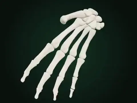
Combined palm support / Palmaris brevis · BETA
About this resource
Supported native palm and finger skeleton; free cutaneous boundary. 12healthy adults,11men/1woman,27±4years; bilateral ultrasound during maximum fifth-digit abduction, supported supinated palm. Published mean relative architecture is adapted to the original source long/thickness axes. Triggering digit abduction, skin dimpling, measured fibers, force and patient limits are not simulated. The native Right source is reflected once for the Left teaching side. Source-end shells and native principal axes are authored geometric references, not measured fiber or enthesis segmentation. The original aponeurosis attachment stays held; the genuine cutaneous-end boundary moves freely. Width compensation is authored, and no skin/fat-pad mesh, contact, force or physiological validation is claimed. Original Palmaris brevis is active while19 native finger muscles stay passive/context; all supported bones and the actual palmar aponeurosis remain held. Its cutaneous end remains a free tissue boundary and is not pinned to a nearby bone. Right/Left use their own recorded32/28% shortening and85/68% thickness references. One actual bone timeline drives every original source body and shared owner. Activity is authored teaching emphasis, not EMG, force, tendon strain or physiological recruitment. Antagonists and other passive context have zero activity; passive lengthening is not automatically eccentric contraction. The selected group remains active during the authored controlled return. Native source identity and exact Rest/reset are retained. BETA teaching approximation; no clinical or faculty validation is claimed.
Inspect the selected source model and its visible limitations.
Model & sources
simLAB · original anatomy teaching source
Source-owned teaching illustration, not a clinical measurement. See the player for its scope and sources.