
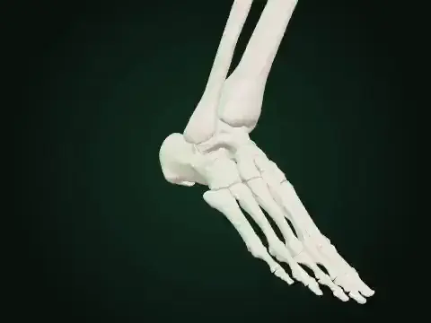
Tibialis posterior · BETA
About this resource
AMA protocol: prone knee flexed90° and ankle slightly dorsiflexed. The model authors a held5° ankle dorsiflexion; the paper does not prescribe that exact angle. The full passive reference does not imply one muscle independently generates or shortens across the entire arc. The achieved posterior heel-line projection reaches the AMA endpoint. A separately solved rotation about the existing oblique subtalar axis carries the distal foot; a source-vertical heel line represents relaxed neutral, not a measured individual calcaneal bisector. The original anterior/malleolar/plantar source course and actual distal bone owners remain. Fixed axes and geometric skinning do not solve translation, contact, tendon sliding, strain, force or loaded arch mechanics. BETA teaching approximation; no clinical or faculty validation is claimed. Activity and geometric readouts are not muscle-force or patient measurements.
Inspect the selected source model and its visible limitations.
Model & sources
simLAB · original anatomy teaching source
Source-owned teaching illustration, not a clinical measurement. See the player for its scope and sources.